The Digestive System - Design: parts of the digestive system
The digestive system may be broken into two parts: a long, winding, muscular tube accompanied by accessory digestive organs and glands. That open-ended tube, known as the alimentary canal or digestive tract, is composed of various organs. These organs are, in order, the mouth, pharynx, esophagus, stomach, small intestine, and large intestine. The rectum and anus form the end of the large intestine. The accessory digestive organs and glands that help in the digestive process include the tongue, teeth, salivary glands, pancreas, liver, and gall bladder.
The walls of the alimentary canal from the esophagus through the large intestine are made up of four tissue layers. The innermost layer is the mucosa, coated with mucus. This protects the alimentary canal from chemicals and enzymes (proteins that speed up the rate of chemical reactions) that break down food and from germs and parasites that might be in that food. Around the mucosa is the submucosa, which contains blood vessels, nerves, and lymph vessels. Wrapped around the submucosa are two layers of muscles that help move food along the canal. The outermost layer, the serosa, is moist, fibrous tissue that protects the alimentary canal and helps it move against the surrounding organs in the body.
The mouth
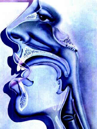
Food enters the body through the mouth, or oral cavity. The lips form and protect the opening of the mouth, the cheeks form its sides, the tongue forms its floor, and the hard and soft palates form its roof. The hard palate is at the front; the soft palate is in the rear. Attached to the soft palate is a fleshy, fingerlike projection called the uvula (from the Latin word meaning "little grape"). Two U-shaped rows of teeth line the mouth—one above and one below. Three pair of salivary glands open at various points into the mouth.
- Alimentary canal (al-i-MEN-tah-ree ka-NAL):
- Also known as the digestive tract, the series of muscular structures through which food passes while being converted to nutrients and waste products; includes the oral cavity, pharynx, esophagus, stomach, large intestine, and small intestine.
- Amylase (am-i-LACE):
- Any of various digestive enzymes that convert starches to sugars.
- Appendix (ah-PEN-dix):
- Small, apparently useless organ extending from the cecum.
- Bile:
- Greenish yellow liquid produced by the liver that neutralizes acids and emulsifies fats in the duodenum.
- Bolus (BO-lus):
- Rounded mass of food prepared by the mouth for swallowing.
- Cecum (SEE-kum):
- Blind pouch at the beginning of the large intestine.
- Chyle (KILE):
- Thick, whitish liquid consisting of lymph and tiny fat globules absorbed from the small intestine during digestion.
- Chyme (KIME):
- Soupylike mixture of partially digested food and stomach secretions.
- Colon (KOH-lun):
- Largest region of the large intestine, divided into four sections: ascending, transverse, descending, and sigmoid (colon is sometimes used to describe the entire large intestine).
- Colostomy (kuh-LAS-tuh-mee):
- Surgical procedure where a portion of the large intestine is brought through the abdominal wall and attached to a bag to collect feces.
- Defecation (def-e-KAY-shun):
- Elimination of feces from the large intestine through the anus.
- Dentin (DEN-tin):
- Bonelike material underneath the enamel of teeth, forming the main part.
- Duodenum (doo-o-DEE-num or doo-AH-de-num):
- First section of the small intestine.
- Emulsify (e-MULL-si-fie):
- To break down large fat globules into smaller droplets that stay suspended in water.
- Enamel (e-NAM-el):
- Whitish, hard, glossy outer layer of teeth.
- Enzymes (EN-zimes):
- Proteins that speed up the rate of chemical reactions.
- Epiglottis (ep-i-GLAH-tis):
- Flaplike piece of tissue at the top of the larynx that covers its opening when swallowing is occurring.
- Esophagus (e-SOF-ah-gus):
- Muscular tube connecting the pharynx and stomach.
- Feces (FEE-seez):
- Solid body wastes formed in the large intestine.
- Flatus (FLAY-tus):
- Gas generated by bacteria in the large intestine.
- Gastric juice (GAS-trick JOOSE):
- Secretion of the gastric glands of the stomach, containing hydrochloric acid, pepsin, and mucus.
- Ileocecal valve (ill-ee-oh-SEE-kal VALV):
- Sphincter or ring of muscule that controls the flow of chyme from the ileum to the large intestine.
- Ileum (ILL-ee-um):
- Final section of the small intestine.
- Jejunum (je-JOO-num):
- Middle section of the small intestine.
- Lacteals (LAK-tee-als):
- Specialized lymph capillaries in the villi of the small intestine.
- Larynx (LAR-ingks):
- Organ between the pharynx and trachea that contains the vocal cords.
- Lipase (LIE-pace):
- Digestive enzyme that converts lipids (fats) into fatty acids.
- Lower esophageal sphincter (LOW-er i-sof-ah-GEE-alSFINGK-ter):
- Strong ring of muscle at the base of the esophagus that contracts to prevent stomach contents from moving back into the esophagus.
- Palate (PAL-uht):
- Roof of the mouth, divided into hard and soft portions, that separates the mouth from the nasal cavities.
- Papillae (pah-PILL-ee):
- Small projections on the upper surface of the tongue that contain taste buds.
- Peristalsis (per-i-STALL-sis):
- Series of wavelike muscular contractions that move material in one direction through a hollow organ.
- Pharynx (FAR-inks):
- Short, muscular tube extending from the mouth and nasal cavities to the trachea and esophagus.
- Plaque (PLACK):
- Sticky, whitish film on teeth formed by a protein in saliva and sugary substances in the mouth.
- Pyloric sphincter (pie-LOR-ick SFINGK-ter):
- Strong ring of muscle at the junction of the stomach and the small intestine that regulates the flow of material between them.
- Rugae (ROO-jee):
- Folds of the inner mucous membrane of organs, such as the stomach, that allow those organs to expand.
- Trypsin (TRIP-sin):
- Digestive enzyme that converts proteins into amino acids; inactive form is trypsinogen.
- Uvula (U-vue-lah):
- Fleshy projection hanging from the soft palate that raises to close off the nasal passages during swallowing.
- Vestigial organ (ves-TIJ-ee-al OR-gan):
- Organ that is reduced in size and function when compared with that of evolutionary ancestors.
- Villi (VILL-eye):
- Tiny, fingerlike projections on the inner lining of the small intestine that increase the rate of nutrient absorption by greatly increasing the intestine's surface area.
THE TONGUE. The muscular tongue is attached to the base of the mouth by a fold of mucous membrane. On the upper surface of the tongue are small projections called papillae, many of which contain taste buds (for a discussion of taste, see chapter 12). Most of the tongue lies within the mouth, but its base extends into the pharynx. Located at the base of the tongue are the lingual tonsils, small masses of lymphatic tissue that serve to prevent infection.
TEETH. Humans have two sets of teeth: deciduous and permanent. The deciduous teeth (also known as baby or milk teeth) start to erupt through the gums in the mouth when a child is about six months old. By the age of two, the full set of twenty teeth has developed. Between the ages of six and twelve, the roots of these teeth are reabsorbed into the body and the teeth begin to fall out. They are quickly replaced by the thirty-two permanent adult teeth. (The third molars, the wisdom teeth, may not erupt because of inadequate space in the jaw. In such cases, they become impacted or embedded in the jawbone and must be removed surgically.)
Teeth are classified according to shape and function. Incisors, the chisel-shaped front teeth, are used for cutting. Cuspids or canines, the pointed teeth next to the incisors, are used for tearing or piercing. Bicuspids (or premolars) and molars, the back teeth with flattened tops and rounded, raised tips, are used for grinding.
Each tooth consists of two major portions: the crown and the root. The crown is the exposed part of the tooth above the gum line; the root is enclosed in a socket in the jaw. The outermost layer of the crown is the whitish enamel. Made mainly of calcium, enamel is the hardest substance in the body.

Underneath the enamel is a yellowish, bonelike material called dentin. It forms the bulk of the tooth. Within the dentin is the pulp cavity, which receives blood vessels and nerves through a narrow root canal at the base of the tooth.
THE SALIVARY GLANDS. Three pair of salivary glands produce saliva on a continuous basis to keep the mouth and throat moist. The largest pair, the parotid glands, are located just below and in front of the ears. The next largest pair, the submaxillary or submandibular glands, are located in the lower jaw. The smallest pair, the sublingual glands, are located under the tongue.

Ivan Petrovich Pavlov (1849–1936) was a Russian physiologist (a person who studies the physical and chemical processes of living organisms) who conducted pioneering research into the digestive activities of mammals. His now-famous experiments with a dog ("Pavlov's dog") to show how the central nervous system affects digestion earned him the Nobel Prize for Medicine or Physiology in 1904.
Interested in the actions of digestion and gland secretion, Pavlov set up an ingenious experiment. In a laboratory, he severed a dog's throat (Pavlov was a skillful surgeon and the animal was unharmed). When the dog ate food, the food dropped out of the animal's throat before reaching its stomach. Through this simulated feeding, Pavlov discovered that the sight, smell, and swallowing of food was enough to cause the secretion of gastric juice. He demonstrated that the stimulation of the vagus nerve (one of the major nerves of the brain) influences the actions of the gastric glands.
In another famous study, Pavlov set out to determine whether he could turn unconditioned (naturally occurring) reflexes or responses of the central nervous system into conditioned (learned) reflexes. He had noticed that laboratory dogs would sometimes salivate merely at the approach of lab assistants who fed them. Pavlov then decided to ring a bell each time a dog was given food. After a while, he rang the bell without feeding the dog. He discovered that the dog salivated at the sound of the bell, even though food was not present. Through this experiment, Pavlov demonstrated that unconditioned reflexes (salivation and gastric activity) could become conditioned reflexes that were triggered by a stimulus (the bell) that previously had no connection with the event (eating).
Ducts or tiny tubes carry saliva from these glands into the mouth. Ducts from the parotid glands open into the upper portion of the mouth; ducts from the submaxillary and sublingual glands open into the mouth beneath the tongue.
The salivary glands are controlled by the autonomic nervous system, a division of the nervous system that functions involuntarily (meaning the processes it controls occur without conscious effort on the part of an individual). The glands produce between 1.1 and 1.6 quarts (1 and 1.5 liters) of saliva each day. Although the flow is continuous, the amount varies. Food (or anything else) in the mouth increases the amount produced. Even the sight or smell of food will increase the flow.
Saliva is mostly water (about 99 percent), with waste products, antibodies, and enzymes making up the small remaining portion. At mealtimes, saliva contains large quantities of digestive enzymes that help break down food. Saliva also controls the temperature of food (cooling it down or warming it up), cleans surfaces in the mouth, and kills certain bacteria present in the mouth.
The pharynx
The pharynx, or throat, is a short, muscular tube extending about 5 inches (12.7 centimeters) from the mouth and nasal cavities to the esophagus and trachea (windpipe). It serves two separate systems: the digestive system (by allowing the passage of solid food and liquids) and the respiratory system (by allowing the passage of air).
The esophagus
The esophagus, sometimes referred to as the gullet, is the muscular tube connecting the pharynx and stomach. It is approximately 10 inches (25 centimeters) in length and 1 inch (2.5 centimeters) in diameter. In the thorax (area of the body between the neck and the abdomen), the esophagus lies behind the trachea. At the base of the esophagus, where it connects with the stomach, is a strong ring of muscle called the lower esophageal sphincter. Normally, this circular muscle is contracted, preventing contents in the stomach from moving back into the esophagus.
The stomach
The stomach is located on the left side of the abdominal cavity just under the diaphragm (a membrane of muscle separating the chest cavity from the abdominal cavity). When empty, the stomach is shaped like the letter J and its inner walls are drawn up into long, soft folds called rugae. When the stomach expands, the rugae flatten out and disappear. This allows the average adult stomach to hold as much as 1.6 quarts (1.5 liters) of material.
The dome-shaped portion of the stomach to the left of the lower esophageal sphincter is the fundus. The large central portion of the stomach is the body. The part of the stomach connected to the small intestine (the curve of the J) is the pylorus. The pyloric sphincter is a muscular ring that regulates the flow of material from the stomach into the small intestine by variously opening and contracting. That material, a soupylike mixture of partially digested food and stomach secretions, is called chyme.

The stomach wall contains three layers of smooth muscle. These layers contract in a regular rhythm—usually three contractions per minute—to mix and churn stomach contents. Mucous membrane lines the stomach. Mucus, the thick, gooey liquid produced by the cells of that membrane, helps protect the stomach from its own secretions. Those secretions—acids and enzymes—enter the stomach through millions of shallow pits that open onto the surface of the inner stomach. Called gastric pits, these openings lead to gastric glands, which secrete about 1.6 quarts (1.5 liters) of gastric juice each day.
Gastric juice contains hydrochloric acid and pepsin. Pepsin is an enzyme that breaks down proteins; hydrochloric acid kills microorganisms and breaks down cell walls and connective tissue in food. The acid is strong enough to burn a hole in carpet, yet the mucus produced by the mucous membrane prevents it from dissolving the lining of the stomach. Even so, the cells of the mucous membrane wear out quickly: the entire stomach lining is replaced every three days. Mucus also aids in digestion by keeping food moist.
William Beaumont (1785–1853) was an American surgeon who served as an army surgeon during the War of 1812 (1812–15) and at various posts after the war. It was at one of these posts that he saw what perhaps no one before him had seen: the inner workings of the stomach.
In 1882, while serving at Fort Mackinac in northern Michigan, Beaumont was presented with a patient named Alexis St. Martin. The French Canadian trapper, only nineteen at the time, has been accidently shot in the stomach. The bullet had torn a deep chunk out of the left side of St. Martin's lower chest. At first, no one thought he would survive, but amazingly he did. However, his wound never completely healed, leaving a 1 inch-wide (2.5 centimeter-wide) opening. This opening allowed Beaumont to put his finger all the way into St. Martin's stomach.
Beaumont decided to take advantage of opening into St. Martin's side to study human digestion. He started by taking small chunks of food, tying them to a string, then inserting them directly into the young man's stomach. At irregular intervals, he pulled the food out to observe the varying actions of digestion. Later, using a hand-held lens, Beaumont peered into St. Martin's stomach. He observed how the human stomach behaved at various stages of digestion and under differing circumstances.
Beaumont conducted almost 240 experiments on St. Martin. In 1833, he published his findings in Experiments and Observations on the Gastric Juice and the Physiology of Digestion , a book that provided invaluable information on the digestive process.
The small intestine
The small intestine is the body's major digestive organ. Looped and coiled within the abdominal cavity, it extends about 20 feet (6 meters) from the stomach to the large intestine. At its junction with the stomach, it measures about 1.5 inches (4 centimeters) in diameter. By the time it meets the large intestine, its diameter has been reduced to 1 inch (2.5 centimeters). Although much longer than the large intestine, the small intestine is called "small" because its overall diameter is smaller.
The small intestine is divided into three regions or sections. The first section, the duodenum, is the initial 10 inches (25 centimeters) closest to the stomach. Chyme from the stomach and secretions from the pancreas and liver empty into this section. The middle section, the jejunum, measures about 8.2 feet (2.5 meters) in length. Digestion and the absorption of nutrients occurs mainly in the jejunum. The final section, the ileum, is also the longest, measuring about 11 feet (3.4 meters) in length. The ileum ends at the ileocecal valve, a sphincter that controls the flow of chyme from the ileum to the large intestine.
The inner lining of the small intestine is covered with tiny, fingerlike projections called villi (giving it an appearance much like the nap of a plush, soft towel). The villi greatly increase the intestinal surface area available for absorbing digested material. Within each villus (singular for villi) are blood capillaries and a lymph capillary called a lacteal. Digested food molecules are absorbed through the walls of the villus into both the capillaries and the lacteal. At the bases of the villi are openings of intestinal glands, which secrete a watery intestinal juice. This juice contains digestive enzymes that convert food materials into simple nutrients the body can readily use. On average, about 2 quarts (1.8 liters) of intestinal juice are secreted into the small intestine each day.
As with the lining of the stomach, a coating of mucus helps protect the lining of the small intestine. Yet again, the digestive enzymes prove too strong for the delicate cells of that lining. They wear out and are replaced about every two days.
The large intestine
Extending from the end of the small intestine to the anus, the large intestine measures about 5 feet (1.5 meters) in length and 3 inches (7.5 centimeters) in diameter. It almost completely frames the small intestine. The large intestine is divided into three major regions: the cecum, colon, and rectum.
Cecum comes from the Latin word caecum , meaning "blind." Shaped like a rounded pouch, the cecum lies immediately below the area where the ileum empties into the large intestine. Attached to the cecum is the slender, fingerlike appendix, which measures about 3.5 inches (9 centimeters) in the average adult. Composed of lymphatic tissue, the appendix seems to have no function in present-day humans. For that reason, scientists refer to it as a vestigial organ (an organ that is reduced in size and function when compared with that of evolutionary ancestors).

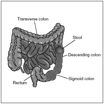
Sometimes used to describe the entire large intestine, the colon is actually the organ's main part. It is divided into four sections: ascending, transverse, descending, and sigmoid. The ascending colon travels from the cecum up the right side of the abdominal cavity until it reaches the liver. It then makes a turn, becoming the transverse colon, which travels horizontally across the abdominal cavity. Near the spleen on the left side, it turns down to form the descending colon. At about where it enters the pelvis, it becomes the S-shaped sigmoid colon.
After curving and recurving, the sigmoid colon empties into the rectum, a fairly straight, 6-inch (15-centimeter) tube ending at the anus, the opening to the outside. Two sphincters (rings of muscle) control the opening and closing of the anus.
Roughly 1.6 quarts (1.5 liters) of watery material enters the large intestine each day. No digestion takes place in the large intestine, only the reabsorption or recovery of water. Mucus produced by the cells in the lining of the large intestine help move the waste material along. As more and more water is removed from that material, it becomes compacted into soft masses called feces. Feces are composed of water, cellulose and other indigestible material, and dead and living bacteria. The remnants of worn red blood cells gives feces their brown color. Only about 3 to 7 ounces (85 to 200 grams) of solid fecal material remains after the large intestine has recovered most of the water. That material is then eliminated through the anus, a process called defecation.
The pancreas
The pancreas is a soft, pink, triangular-shaped gland that measures about 6 inches (15 centimeters) in length. It lies behind the stomach, extending from the curve of the duodenum to the spleen. While a part of the digestive system, the pancreas is also a part of the endocrine system, producing the hormones insulin and glucagon (for a further discussion of this process, see chapter 3).
Primarily a digestive organ, the pancreas produces pancreatic juice that helps break down all three types of complex food molecules in the small intestine. The enzymes contained in that juice include pancreatic amylase, pancreatic lipase, and trypsinogen. Amylase breaks down starches into simple sugars, such as maltose (malt sugar). Lipase breaks down fats into simpler fatty acids and glycerol (an alcohol). Trypsinogen is the inactive form of the enzyme trypsin, which breaks down proteins into amino acids. Trypsin is so powerful that if produced in the pancreas, it would digest the organ itself. To prevent this, the pancreas produces trypsinogen, which is then changed in the duodenum to its active form.
Pancreatic juice is collected from all parts of the pancreas through microscopic ducts. These ducts merge to form larger ducts, which eventually combine to form the main pancreatic duct. This duct, which runs the length of the pancreas, then transports pancreatic juice to the duodenum of the small intestine.
The liver
The largest glandular organ in the body, the liver weighs between 3 and 4 pounds (1.4 and 1.8 kilograms). It lies on the right side of the abdominal cavity just beneath the diaphragm. In this position, it overlies and almost completely covers the stomach. Deep reddish brown in color, the liver is divided into four unequal lobes: two large right and left lobes and two smaller lobes visible only from the back.
The liver is an extremely important organ. Scientists have discovered that it performs over 200 different functions in the body. Among its many functions are processing nutrients, making plasma proteins and blood-clotting chemicals, detoxifying (transforming into less harmful substances) alcohol and drugs, storing vitamins and iron, and producing cholesterol.
One of the liver's main digestive functions is the production of bile. A watery, greenish yellow liquid, bile consists mostly of water, bile salts, cholesterol, and assorted lipids or fats. Liver cells produce roughly 1 quart (1 liter) of bile each day. Bile leaves the liver through the common hepatic duct. This duct unites with the cystic duct from the gall bladder to form the common bile duct, which delivers bile to the duodenum.
In the small intestine, bile salts emulsify fats, breaking them down from large globules into smaller droplets that stay suspended in the watery fluid in the small intestine. Bile salts are not enzymes and, therefore, do not digest fats. By breaking down the fats in to smaller units, bile salts aid the fatdigesting enzymes present in the small intestine.
The gall bladder
The gall bladder is a small, pouchlike, green organ located on the undersurface of the right lobe of the liver. It measures 3 to 4 inches (7.6 to 10 centimeters) in length. The gall bladder's function is to store bile, of which it can hold about 1.2 to 1.7 ounces (35 to 50 milliliters).
The liver continuously produces bile. When digestion is not occurring, bile backs up the cystic duct and enters the gall bladder. While holding the bile, the gall bladder removes water from it, making it more concentrated. When fatty food enters the duodenum once again, the gall bladder is stimulated to contract and spurt out the stored bile.

Comment about this article, ask questions, or add new information about this topic: